
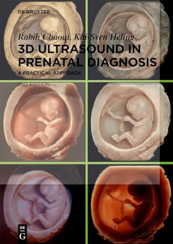
3D Ultrasound in Prenatal Diagnosis A Practical Approach 2nd Edition by Rabih Chaoui, Kai Sven Heling ISBN 3111249093 9783111249094


Original price was: $50.00.$35.00Current price is: $35.00.
3D Ultrasound in Prenatal Diagnosis A Practical Approach 2nd Edition by Rabih Chaoui, Kai Sven Heling – Ebook PDF Instant Download/Delivery: 3111249093, 9783111249094
Full download 3D Ultrasound in Prenatal Diagnosis A Practical Approach 2nd Edition after payment
Product details:
ISBN 10: 3111249093
ISBN 13: 9783111249094
Author: Rabih Chaoui, Kai Sven Heling
3D Ultrasound in Prenatal Diagnosis A Practical Approach 2nd Table of contents:
Part I: Basics of 3D Sonography
1 Basics of 3D and 4D Volume Acquisition
1.1 Introduction
1.2 Preparing volume acquisition
1.3 Transducers for 3D/4D acquisition
1.4 Types of volume acquisition
1.5 Display of a volume data set
1.6 Conclusions
2 Orientation and Navigation within a Volume Data set
2.1 Introduction
2.2 Storing and exporting volume data sets
2.3 Orientation in the three orthogonal planes
2.4 Navigation within the orthogonal planes
2.5 Artifacts in the multiplanar mode
2.6 Conclusions
Part II: 3D Display Methods
3 3D Rendering and the Rendering Modes
3.1 Introduction
3.2 The render box and the orientation within a 3D volume
3.3 Artifacts in 3D rendering
3.4 Different rendering modes and mixture of modes
3.5 Special effects in 3D: dynamic depth rendering and light source
3.6 Threshold, transparency, brightness, and color scales
3.7 Magicut, the electronic scalpel
3.8 SonoRender live
3.9 Conclusions
4 Volume Contrast Imaging (VCI)
4.1 Introduction
4.2 Principle of VCI
4.3 Static VCI
4.4 VCI with color Doppler
4.5 4D with VCI-Omniview
4.6 4D with VCI-A
4.7 Conclusions
5 Multiplanar Display I: Orthogonal Mode and Omniview Planes
5.1 Principle
5.2 Multiplanar reconstruction and different displays of cross-sectional images
5.3 Practical approach in orthogonal mode
5.4 Practical approach in obtaining an “anyplane” using Omniview mode
5.5 Typical applications of the Omniview mode
5.6 Conclusions
6 Multiplanar Display II: The Tomography Mode
6.1 Principle
6.2 Practical approach
6.3 Typical applications of the tomography mode
6.4 Conclusions
7 The Surface Mode
7.1 Principle
7.2 Practical approach
7.3 Typical applications of the surface mode
7.4 Conclusions
8 The Maximum Mode
8.1 Principle
8.2 Practical approach
8.3 Typical applications for bones’ visualization
8.4 Conclusions
9 The Minimum Mode
9.1 Principle
9.2 Practical approach
9.3 Typical applications of the minimum mode
9.4 Conclusions
10 The Inversion Mode
10.1 Principle
10.2 Practical approach
10.3 Typical applications of the inversion mode
10.4 Conclusions
11 The Silhouette Mode
11.1 Principle
11.2 Practical approach
11.3 Typical applications of the silhouette mode
11.4 Conclusions
12 The Glass-Body Mode
12.1 Principle
12.2 Practical approach
12.3 Typical applications of the glass-body mode
12.4 Conclusions
13 Calculation of 3D Volumes
13.1 Principle
13.2 Practical approach
13.3 Typical applications of volume calculations
13.4 Conclusions
14 The Electronic Matrix Transducer
14.1 Principle
14.2 Biplane display
14.3 Volume Contrast Imaging of the A-plane (VCI-A)
14.4 The new tool VCI-2D
14.5 Conclusions
Part III: Clinical Applications in Prenatal Diagnosis
15 The Fetal Face in 3D
15.1 Examination of the face in 2D and 3D ultrasound
15.2 The normal face in 3D surface mode
15.3 The normal face in 4D surface mode
15.4 The normal face at different gestational ages
15.5 The abnormal face in 3D/4D
15.6 The normal and abnormal ear
15.7 The face in multiplanar display
15.8 Conclusions
16 3D Fetal Neurosonography
16.1 Introduction
16.2 Transabdominal 3D examination of the brain and multiplanar reconstruction
16.3 Transvaginal 3D examination of the brain and multiplanar reconstruction
16.4 Fetal brain visualized with 3D volume rendering
16.5 The intracerebral vascular system in 3D glass-body mode
16.6 3D of the fetal brain before 14 gestational weeks
16.7 Conclusions
17 3D of Skull, Spine, and Limbs
17.1 Examination of the fetal skeleton with 2D ultrasound
17.2 3D imaging of the facial and cranial bones
17.3 3D imaging of the fetal spine and ribs
17.4 3D imaging of the fetal limbs
17.5 Conclusions
18 3D of Intrathoracic and Abdominal Organs
18.1 Introduction
18.2 The intrathoracic organs
18.3 The gastrointestinal organs
18.4 The urogenital system
18.5 Conclusions
19 Fetal Echocardiography in 3D and STIC
19.1 Fetal cardiac scanning in grayscale and color Doppler
19.2 Different acquisitions of cardiac volume data sets
19.3 Fetal heart in 3D and STIC with multiplanar reconstruction
19.4 Fetal heart in grayscale STIC with rendering
19.5 Fetal heart in color Doppler STIC with glass-body rendering
19.6 Automated cardiac volume evaluation and Sono-VCAD
19.7 Other applications of 3D/4D fetal heart examination
19.8 Conclusions
20 3D in Early Pregnancy
20.1 Introduction
20.2 3D volume rendering in early gestation
20.3 Multiplanar display in early gestation
20.4 3D Color Doppler in early gestation
20.5 Conclusions
21 Further literature references and sources
People also search for 3D Ultrasound in Prenatal Diagnosis A Practical Approach 2nd:
3d ultrasound in prenatal diagnosis a practical approach
3d ultrasound in prenatal diagnosis a practical approach 2nd edition
3d prenatal ultrasound near me
do you get 3d ultrasound during pregnancy
what is 3d ultrasound for pregnancy
Tags: Rabih Chaoui, Kai Sven Heling, 3D Ultrasound, Prenatal Diagnosis